At CDN, we love diving into the science, but just as importantly, we love getting to know the scientists behind the discoveries. In this interview, we spoke with Jeff (PI) and Ari (Graduate Student) about their backgrounds, the journey that led them to their current research, and the recent paper on bacterial MERFISH—a groundbreaking technology expanding our ability to map bacterial RNA organization in single cells.
Meet the Scientists
Ari: A Journey from Switzerland to Boston
Ari grew up in Geneva, Switzerland, where he pursued his studies before making his way to Harvard Medical School for a fellowship during his master’s at EPFL. He joined Jeff’s lab, a lab deeply embedded in bacterial research, after the pandemic. This was about a year into his PhD program, following completion of his master’s project in a different lab.
What drives Ari? The thrill of discovery.
“We can’t discover new continents anymore—that’s been done. Maybe one day, we’ll explore other planets, but for now, biology is still an open frontier,” he says. “Problem-solving and troubleshooting can be frustrating, but they’re also what make this job so fun. When something doesn’t work, I get to ask: Why didn’t it work? How can I test it? How can I measure it?”
Jeff: From Physics to Biology
Jeff’s path took him from physics to biology. He earned his PhD at UC Berkeley, where he developed optical tweezers—biological “tractor beams” that allowed researchers to manipulate single molecules and study enzymes interacting with nucleic acids.
Wanting to explore the complexities of the cell, he transitioned to bacterial research during his postdoc. His work led to an exciting collaboration that helped pioneer spatial biology using a multiplexed RNA imaging method, MERFISH. “Our goal is to develop technologies that let us see things that were simply unobservable before,” Jeff explains.
Bacterial MERFISH: A New Window into Microbial Life
So how did this project come about?
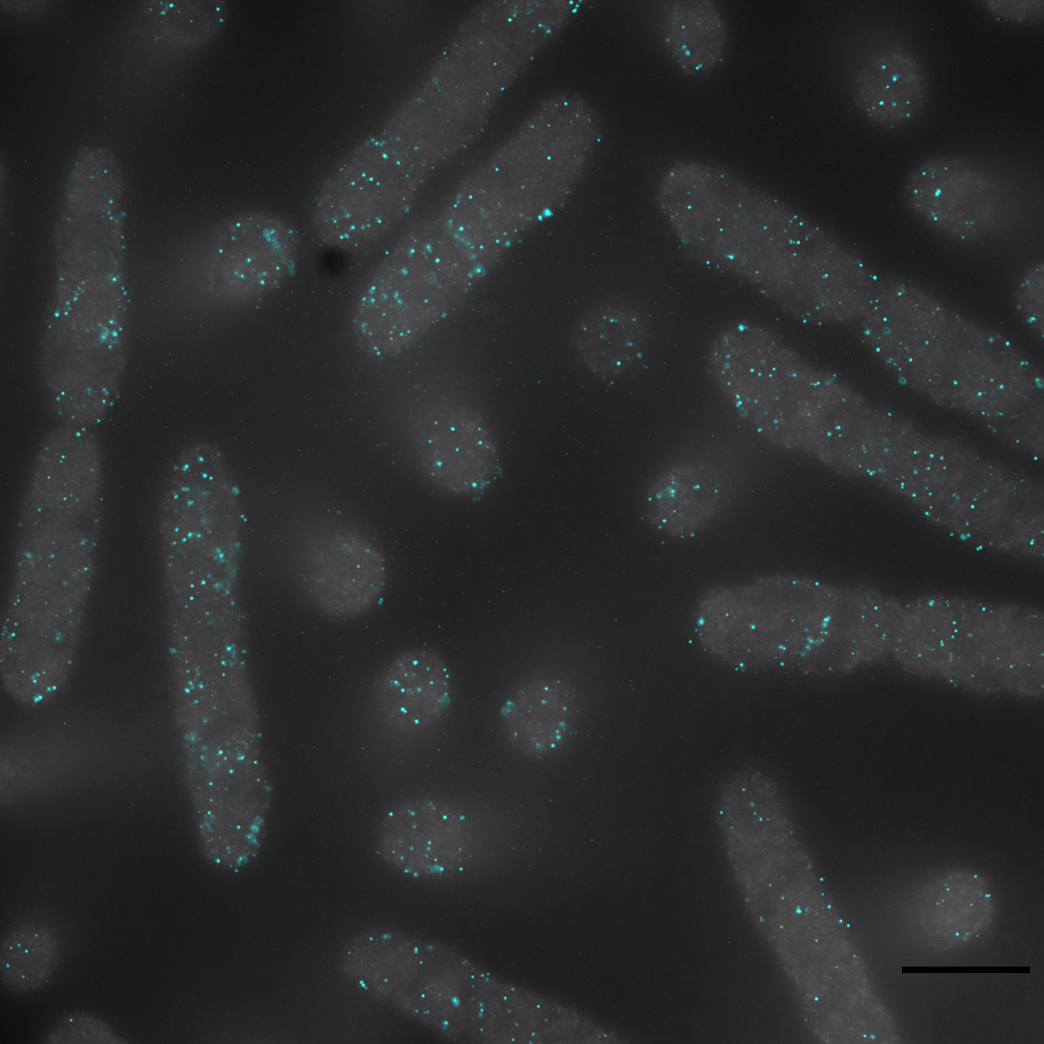
MERFISH had already transformed how scientists study tissue biology, but Jeff wanted to apply it to bacteria. The problem? Bacteria are tiny, and their RNA is packed far more densely than in mammalian cells—about 1,000 times denser. This made it impossible to distinguish individual RNA molecules with traditional microscopy.
Ari and his collaborator, Dr. Yuanyou Wang, took on the challenge. “We had to solve a three-order-of-magnitude RNA density problem before we could even get started,” Jeff says.
Expanding Bacteria—Literally
To overcome this, the team turned to expansion microscopy, a technique that physically enlarges cells using hydrogels. Imagine a balloon: if you write on it and then inflate it, the text spreads out, becoming easier to read. Expansion microscopy works the same way, allowing researchers to visualize bacterial RNA organization at unprecedented resolution.
“Instead of making the microscope better, we make the sample bigger,” Ari explains. “We embed bacteria in a hydrogel, then let it expand. The molecules are pulled away from each other but are not themselves stretched that much, making them resolvable under a standard microscope.”
Mapping Bacterial RNA Organization
With this approach, Ari and his team were able to do something that had never been done before: map the spatial organization of over half of the E. coli transcriptome.
For years, scientists assumed bacteria had little RNA organization—after all, their small size and rapid molecular diffusion suggested otherwise. But Ari’s work contributed to a growing appreciation that bacterial transcriptomes are organized within the cell.
. “We found a surprising diversity of RNA spatial patterns,” Jeff says. “Some RNAs are evenly distributed, but many exhibit strikingly distinct localizations.”
These findings raise new questions: Why do bacteria organize their RNAs? How does this organization influence cellular function? The research has opened an entirely new avenue for studying bacterial cell biology.
Unexpected Insights into Bacterial Metabolism
One of the most surprising findings came when the team looked at how bacteria adapt to different nutrients. Bacteria don’t just switch to metabolizing a new sugar when their preferred one runs out. Instead, they go through an exploratory transcriptional phase, testing for alternative sugars—even ones that aren’t present in the environment.
“It’s like they’re sending out molecular feelers,” Ari explains. “They don’t immediately switch to the second-best sugar. Instead, they express genes for several different metabolic pathways before committing.”
Using their technology, the team was also able to uncover the molecular activities of subpopulations within identical bacterial cultures—some focused on amino acid synthesis, others on their functional machinery.
“This is a fundamental insight into bacterial gene regulation,” Jeff adds. “Even in minimal conditions, bacteria are exhibiting metabolic diversity.”
The Future of Bacterial MERFISH
Beyond understanding bacterial behavior in the lab, this technology has potential real-world applications. For the first time, researchers can apply MERFISH to bacteria inside living hosts, providing unprecedented insight into microbial interactions in their natural environments.
“We don’t just want to study bacteria in test tubes—we want to understand them in their real-world settings,” Ari says. “This technique gives us a way to do that.”
Closing Thoughts
Before wrapping up, we asked Jeff and Ari a few fun questions.
What’s the most impactful scientific advancement of the last decade?
For Jeff, it’s the explosion of spatial transcriptomics—merging genomics with microscopy to study biology at an unprecedented resolution. “The ability to map tens of thousands of RNAs while preserving spatial context is a game-changer,” he says.
Ari, on the other hand, struggles to pick just one. “AI is changing the way we process biological data. Gene therapy is becoming a reality. And cryo-electron microscopy has revolutionized how we visualize molecular structures. There’s too much to choose from!”
If you weren’t a scientist, what would you be doing?
Jeff: “I love discovery, but I also love mentorship and teaching. If I had to drop research, I’d probably still be a teacher.”
Ari: “I love the engineering side of research—building tools, optimizing processes. If I weren’t in science, I’d probably go into industrial design or automation.”
Final Takeaways
Jeff and Ari’s work is pushing the boundaries of bacterial research, making it possible to study microbial gene expression with an unprecedented level of detail.
Their study demonstrates that bacterial RNA isn’t randomly distributed—it’s spatially organized, and understanding this could transform how we think about bacterial physiology. By adapting MERFISH for bacteria, they’ve unlocked new opportunities to study bacterial adaptation, gene regulation, and cellular behavior in ways that were previously impossible.
The future of bacterial spatial transcriptomics is just beginning, and we can’t wait to see what comes next.